
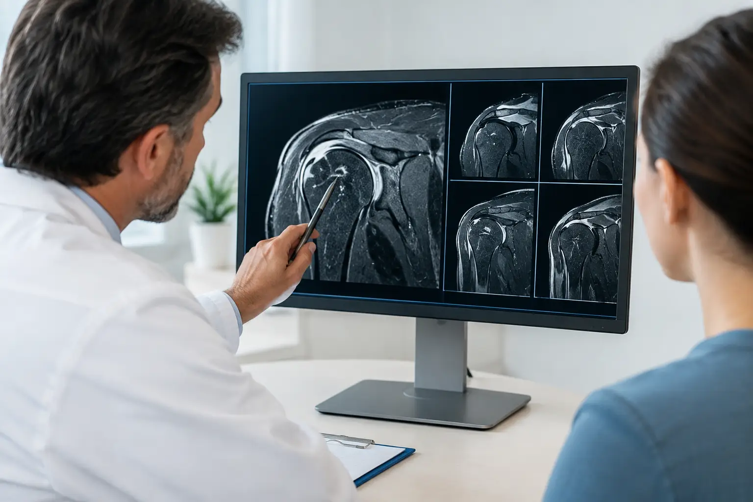
A shoulder MRI report can feel alarming when you open it after a painful fall, a dislocation, or weeks of lost velocity and strength. Terms such as “tear,” “degeneration,” and “edema” can sound definitive. Reading shoulder MRI results is useful, but the report is only one part of the diagnosis. Your symptoms, physical examination, sport, age, injury mechanism, and goals all matter when deciding what treatment will best restore safe function.
For an athlete, the central question is rarely just “What does the MRI show?” It is whether the finding explains the pain or instability, whether it is likely to worsen with continued play, and what path offers the best chance of returning to sport without taking unnecessary risks.
What a Shoulder MRI Can Show
MRI uses magnetic imaging to evaluate soft tissues that may not be visible on an X-ray. In the shoulder, that includes the rotator cuff tendons, labrum, biceps tendon, cartilage, joint capsule, ligaments, bursa, and bone marrow. It can also show fluid, inflammation, swelling after injury, and some fractures that are difficult to see on routine X-rays.
Most reports begin with a technical description, followed by observations about each structure and an “Impression” section. The impression is the radiologist’s concise list of the most significant findings. It is often the first place patients look, but it should not be read as a treatment plan.
An MRI is highly valuable, yet it has limits. A scan is taken with the shoulder still, while many athletic symptoms happen during throwing, lifting, tackling, serving, or reaching overhead. Some tears are clinically meaningful; others may be longstanding changes that are not causing symptoms. A shoulder specialist combines the images with an examination to determine which finding deserves attention.
Reading Shoulder MRI Results: Common Terms
Rotator cuff tendinosis, partial tear, and full-thickness tear
The rotator cuff is a group of four tendons that helps center the ball of the upper arm in the shoulder socket and control lifting and rotation. “Tendinosis” means chronic tendon wear or degeneration. It does not necessarily mean a tendon is torn, and many patients improve with targeted physical therapy, activity modification, and appropriate nonoperative care.
A “partial-thickness tear” involves only part of the tendon. Reports may describe its depth, location, and whether it is on the joint side or bursal side of the tendon. Management depends on pain, weakness, sport demands, tear size, and response to rehabilitation.
A “full-thickness” or “complete” tear extends through the tendon. The report may also mention retraction, meaning the torn tendon has pulled away from its normal attachment, and muscle atrophy or fatty changes. These details can affect the timing and complexity of repair. In an active patient with a traumatic tear and substantial weakness, timely specialist evaluation is especially important.
Labral tears, SLAP tears, and Bankart lesions
The labrum is a rim of cartilage around the socket that deepens the joint and contributes to stability. Labral injuries are common after dislocations, falls, repetitive overhead activity, and contact sports.
A SLAP tear involves the upper portion of the labrum, near the attachment of the biceps tendon. It can cause deep shoulder pain, clicking, or reduced performance in throwers, but MRI findings must be interpreted carefully. Some superior labral changes are seen in athletes and adults without causing symptoms.
A Bankart lesion usually refers to a tear of the lower front portion of the labrum after an anterior shoulder dislocation. If a report also identifies bone loss from the front of the socket or a Hill-Sachs lesion on the humeral head, it may indicate a more significant instability event. For athletes who have recurrent dislocations or feel the shoulder slipping, these findings help guide discussions about stabilization and return-to-play risk.
Biceps tendon findings
The long head of the biceps tendon travels through the shoulder joint and attaches near the top of the labrum. MRI reports may describe biceps tendinitis, tenosynovitis, subluxation, or a tear. “Subluxation” means the tendon is shifting out of its normal groove, often alongside injury to nearby rotator cuff tissue.
Biceps findings can be a genuine pain source, particularly with front-of-shoulder pain during lifting or throwing. They can also appear alongside other issues, so treatment is based on the complete clinical picture rather than one line in the report.
Bursitis, impingement, and AC joint arthritis
The bursa is a thin fluid-filled sac that reduces friction between the rotator cuff and surrounding structures. “Subacromial-subdeltoid bursitis” means inflammation or extra fluid in that space. It may accompany a cuff problem, repetitive overhead loading, or irritation from altered shoulder mechanics.
Reports may also mention acromioclavicular, or AC, joint arthritis. This is wear at the small joint on top of the shoulder, near the collarbone. It can be painful after a direct impact or with cross-body movements, but mild AC joint arthritis is also common and may not be responsible for a patient’s symptoms.
The word “impingement” is sometimes used to describe narrowing above the rotator cuff. It is not a complete diagnosis by itself. The shape of the acromion, posture, strength, scapular control, and tendon health may all contribute to pain with overhead activity.
Bone marrow edema, cysts, and cartilage changes
Bone marrow edema means increased fluid signal inside a bone. After an acute injury, it may represent a bone bruise. It can also occur with stress-related injury, arthritis, or inflammation. Its meaning depends on where it is located and whether there was a specific trauma.
Cartilage loss, chondral wear, or osteoarthritis refers to changes in the smooth joint surface. A small cartilage defect may be managed very differently in a 16-year-old collision athlete than in an adult with longstanding pain and stiffness. Cysts may be benign incidental findings, or they may point to a nearby labral tear or chronic joint change. Context is essential.
Why the MRI Report Does Not Make the Decision
A report can identify anatomy, but it cannot measure how your shoulder performs under the demands of your life. A recreational tennis player, a high school quarterback, a collegiate swimmer, and a parent who needs to lift a child may have the same MRI phrase but need different recommendations.
The timing of the injury matters as well. A traumatic event followed by immediate weakness, loss of motion, or instability is different from gradual soreness that developed over a season. The physical examination helps determine whether an MRI finding matches the painful area, weakness pattern, or instability symptoms.
Treatment is not automatically surgery. Many tendon, bursal, and mild labral findings respond well to a structured rehabilitation program that restores motion, rotator cuff strength, scapular control, and sport-specific mechanics. In selected cases, anti-inflammatory measures, an injection, or orthobiologic options may be considered. Surgical repair or reconstruction may be appropriate when there is significant structural damage, recurrent instability, persistent functional limitation, or a high risk of further injury with continued sport.
When to Arrange a Prompt Shoulder Evaluation
Do not wait to seek care if the shoulder looks deformed after an injury, you cannot lift the arm, you have numbness or a cold hand, or you believe the joint has dislocated. These symptoms may require urgent evaluation.
A timely sports medicine or orthopaedic consultation is also wise when an MRI follows a traumatic injury and shows a full-thickness rotator cuff tear, substantial tendon retraction, a labral tear after dislocation, or bone loss related to instability. Repeated episodes of the shoulder slipping out, nighttime pain that is not improving, and ongoing weakness despite rest are also reasons to be evaluated.
For young athletes, growth plates and developing anatomy add another layer of interpretation. Pediatric and adolescent shoulders should be assessed with age, sport exposure, and future participation in mind rather than treated as smaller adult shoulders.
Bring the Images, Not Only the Report
At your appointment, bring the actual MRI images on disc or through the imaging center’s electronic access system, along with the written report. A specialist can review the images directly and compare them with your examination. It also helps to write down when symptoms started, what movement causes pain, whether there was a pop or dislocation, prior treatment, and your sport calendar.
Useful questions include whether the MRI finding matches your symptoms, what activities are safe now, whether physical therapy is appropriate, and what benchmarks should be met before returning to practice or competition. If surgery is being discussed, ask what problem it is intended to correct, the expected rehabilitation timeline, and what return-to-sport criteria will be used.
A shoulder MRI can provide clarity, but the best next step comes from interpreting the scan in the context of the person who needs to use that shoulder. With an accurate diagnosis and an individualized plan, the focus can move from worrying about report language to rebuilding strength, stability, and confidence for the activities that matter most.
Leave a Reply