
A pitcher feels a sharp catch in the shoulder during a throw. A soccer player develops knee swelling after a pivot. A parent wants to know whether their child needs an MRI immediately or if an ultrasound will provide the answer. In the MRI versus ultrasound decision, the best test is not simply the most detailed or most familiar one. It is the study that can answer the clinical question and help direct the next step toward safe recovery.
Both MRI and ultrasound are valuable tools in sports medicine. They do different jobs, have different strengths, and are often selected after a thorough history and physical examination. Imaging should support clinical judgment, not replace it. The location of pain, the movement that caused it, the examination findings, and the athlete’s goals all affect which test is appropriate.
MRI Versus Ultrasound: The Core Difference
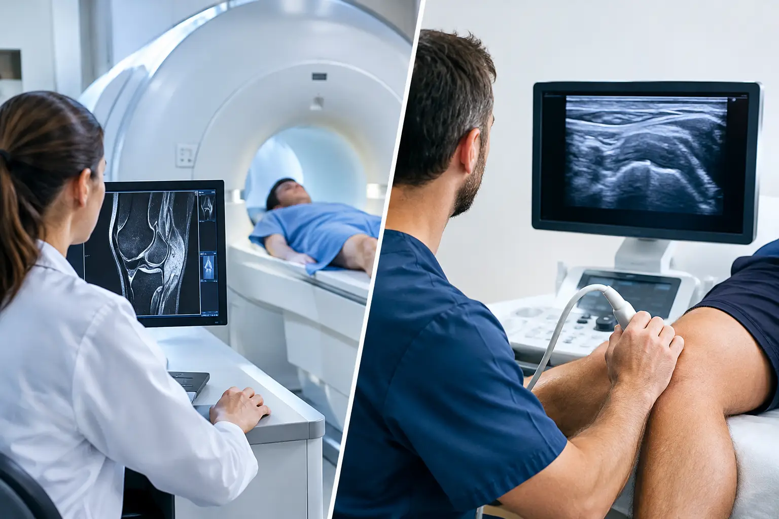
MRI, or magnetic resonance imaging, uses a powerful magnet and radio waves to create detailed images of bones, cartilage, ligaments, tendons, muscles, and other soft tissues. It does not use radiation. MRI is particularly useful when an injury may involve structures deep within a joint or when the diagnosis depends on seeing anatomy that ultrasound cannot reliably reach.
Ultrasound uses high-frequency sound waves to create images in real time. It is especially effective for evaluating many superficial structures, including tendons, muscles, bursae, fluid collections, and some nerves. During an ultrasound examination, the physician may move the joint or have the patient perform a specific motion. That dynamic view can reveal tendon snapping, impingement, instability, or pain that only occurs with movement.
Neither test is universally better. An MRI offers a broader, more detailed look at deep joint structures. Ultrasound offers speed, dynamic assessment, and the ability to compare the painful side with the unaffected side during the same visit. The right choice depends on what the physician suspects and what information will change treatment.
When an MRI Is Usually More Helpful
MRI is often the preferred study when a sports injury may involve internal joint structures. For example, a knee MRI can identify an ACL tear, meniscus tear, cartilage injury, bone bruise, or damage to other ligaments. In an athlete whose knee buckled during a noncontact pivot and then became swollen, MRI can help define the extent of injury and inform decisions about bracing, rehabilitation, or reconstruction.
In the shoulder, MRI is frequently used to evaluate labral tears, rotator cuff tears, cartilage damage, and instability-related injuries. A young athlete with repeated shoulder dislocations, a football player with persistent pain after a collision, or a swimmer with deep shoulder pain may need MRI findings to clarify whether the problem is muscular irritation, tendon injury, labral damage, or another condition inside the joint.
MRI can also be important for injuries that are not visible on standard X-rays. Stress reactions and stress fractures, particularly in the hip, pelvis, lower leg, or foot, may require MRI because it can show bone marrow changes before a fracture becomes obvious. This matters for runners and other athletes who may otherwise continue training through an injury that needs rest and protection.
An MRI examination is painless, but it requires the patient to remain still in a narrow scanner for a period of time. The machine can be loud, and some patients feel anxious in enclosed spaces. Younger children or patients with severe claustrophobia may need additional preparation. MRI also costs more than ultrasound and may require insurance authorization, depending on the plan and clinical circumstances.
When Ultrasound Can Be the Better First Test
Ultrasound is highly useful when symptoms point to a tendon, muscle, bursa, or other structure close to the skin. It can evaluate common problems such as rotator cuff tendinopathy or tears, biceps tendon abnormalities, tennis elbow, Achilles tendon injuries, patellar tendon conditions, muscle strains, and fluid around a joint.
For certain shoulder complaints, ultrasound can provide a fast and meaningful assessment. It can show whether the rotator cuff tendon is thickened, inflamed, partially torn, or fully torn. It can also identify bursitis and assess the biceps tendon while the shoulder moves. However, if the concern is a labral tear or instability inside the shoulder joint, an MRI is generally more informative.
Ultrasound can also be useful in guiding an injection with precision. When an injection is part of a treatment plan, real-time imaging allows the physician to see the needle path and confirm placement in the intended area. This may be helpful for certain joint, tendon sheath, or bursal conditions, including situations where an ortho-biologic treatment such as platelet-rich plasma is being considered.
One practical advantage is convenience. Ultrasound can often be performed in the office, produces immediate images, and does not involve radiation, magnets, or a confined scanner. For an athlete trying to understand a suspected muscle or tendon injury quickly, that efficiency can reduce uncertainty and help begin an appropriate recovery plan.
Ultrasound does have limits. Its accuracy depends substantially on the skill and experience of the person performing and interpreting the study. It does not see through bone well, and deep structures inside the hip, knee, and shoulder are not evaluated as completely as they are with MRI. A normal ultrasound does not necessarily rule out a deep joint injury.
Imaging Is Chosen Based on the Injury Pattern
The same body part can require different imaging depending on the mechanism of injury and examination. Consider shoulder pain. An older active adult who notices gradual weakness and pain when lifting may benefit from ultrasound or MRI to assess the rotator cuff. A teenage baseball player with painful clicking and loss of throwing velocity may need MRI because a labral injury is more strongly suspected. An athlete with a visible shoulder deformity after a fall needs prompt examination and often X-rays first to evaluate for a fracture or dislocation.
The knee is similar. Ultrasound can help assess superficial swelling, tendon injuries, and some muscle problems around the knee. But it is not the primary test for a suspected ACL or meniscus tear. When there is twisting trauma, a pop, rapid swelling, locking, or repeated giving way, MRI is often the more appropriate advanced study.
For pediatric athletes, imaging decisions deserve particular care. Growing bones, growth plates, training demands, and the desire to return for an important season can complicate the picture. The goal is neither to order advanced imaging automatically nor to delay it when the findings could affect treatment. A sports medicine specialist can help families understand what each test may show, what it cannot show, and whether imaging is likely to change the plan.
Why the Physical Examination Still Matters
An image can reveal abnormalities that are unrelated to the athlete’s pain. This is common in active adults, especially in the shoulder, knee, and spine. A scan may show age-related tendon changes or a small cartilage irregularity that does not require surgery. Treating an MRI report without considering the patient can lead to unnecessary worry or care that does not address the real problem.
A careful examination helps connect imaging findings to symptoms. Strength testing, range of motion, stability tests, tenderness, swelling, and sport-specific movements help identify which structures are likely involved. The examination also determines whether an athlete has warning signs that need urgent attention, such as a fracture, infection, loss of circulation, severe weakness, or a locked joint.
At Dr. Louis Rizio’s practice, the imaging discussion is part of a larger treatment decision: what is injured, what level of activity the patient hopes to regain, and what approach offers the safest path forward. Some injuries improve with activity modification and focused physical therapy. Others may need bracing, an injection, or surgical repair. Imaging is most useful when it makes that decision clearer.
What to Ask Before Scheduling a Scan
If imaging has been recommended, ask what specific diagnosis is being considered and how the result could change treatment. It is also reasonable to ask whether X-rays are needed first, whether ultrasound can answer the question, and whether an MRI should include contrast. Most sports injury MRIs do not require contrast, although certain shoulder or postoperative evaluations may call for a specialized study.
Bring prior imaging and reports to the consultation when possible. Comparing an old MRI, X-ray, or operative report with current symptoms can prevent duplicate testing and provide useful context. Athletes should also mention prior surgeries, implanted devices, metal fragments, and any difficulty with confined spaces before an MRI is scheduled.
The most useful scan is the one that leads to a clearer plan, whether that means confidence to begin rehabilitation or the information needed to repair a significant injury. If pain, swelling, instability, weakness, or loss of function is keeping you or your child out of the activities you value, a focused orthopaedic evaluation can help determine the next right step.
Leave a Reply