
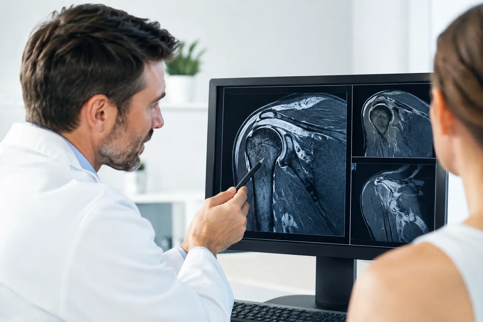
A shoulder MRI report can feel alarming when it includes terms such as tear, degeneration, edema, or labral abnormality. Yet shoulder MRI interpretation is not simply a matter of finding a phrase in the report and deciding whether surgery is needed. For an athlete, active adult, or parent of a young player, the meaningful question is whether the MRI finding explains the pain, instability, weakness, or loss of function that is limiting activity.
Why Shoulder MRI Interpretation Requires Context
MRI is an excellent tool for viewing the soft tissues and bones of the shoulder. It can show the rotator cuff tendons, labrum, cartilage, biceps tendon, joint capsule, ligaments, and bone marrow in much greater detail than a standard X-ray. In many cases, it helps confirm a suspected injury and guides the next step in treatment.
Still, an MRI is one part of the evaluation, not the entire diagnosis. Some changes seen on MRI are common in people who have no shoulder pain, especially as they get older or after years of overhead sports, weight training, or physical work. A partial rotator cuff tear or labral irregularity may be relevant in one patient and incidental in another.
A sports medicine specialist compares the images and report with the injury mechanism, physical examination, symptoms, level of sport, and goals. A pitcher with painful loss of velocity after a throwing injury requires a different interpretation than an active adult with gradual pain while reaching overhead. The image may look similar, but the care plan may not be.
What the MRI Is Looking At
The shoulder is built for range of motion, which also makes it vulnerable to injury. The ball of the upper arm bone sits in a shallow socket called the glenoid. A rim of cartilage called the labrum deepens that socket, while the rotator cuff muscles and tendons help center and move the joint.
Rotator Cuff Tendons
The rotator cuff includes four muscles and their tendons. These tendons can become inflamed, frayed, partially torn, or fully torn. A report may describe tendinosis, which means tendon degeneration or wear, rather than a complete rupture.
A partial-thickness tear does not automatically require surgery. Its significance depends on the depth and location of the tear, the patient’s strength, pain level, response to physical therapy, and activity demands. A full-thickness tear means the tendon is torn all the way through. In a patient with acute weakness after a fall, dislocation, or lifting injury, timely specialist evaluation is particularly important because delayed repair can allow the tendon and muscle to change over time.
MRI can also show tendon retraction and muscle atrophy or fatty change. These findings help a surgeon assess repairability and discuss realistic recovery expectations when surgery is being considered.
Labrum and Shoulder Instability
The labrum may tear after a shoulder dislocation, repetitive throwing, a fall on an outstretched arm, or traction injury. An anterior-inferior labral tear is commonly associated with the shoulder slipping out the front of the joint. A SLAP tear involves the upper labrum near the attachment of the biceps tendon.
Labral findings deserve careful interpretation, particularly in overhead athletes. Some labral changes may reflect normal variation or accumulated stress rather than the source of pain. The examination matters: Does the shoulder feel loose or unstable? Has it dislocated or partially slipped out? Is there painful clicking, dead-arm sensation, or loss of control during throwing?
For a young contact athlete with repeated instability, a structural labral injury may point toward surgical stabilization after an individualized discussion. For another patient, a period of focused rehabilitation may restore strength and control without surgery.
Cartilage, Bone, and the Biceps Tendon
MRI may identify cartilage wear, arthritis, bone bruising, cysts, fractures not visible on X-ray, or changes from prior instability. After a dislocation, for example, the scan may show a Hill-Sachs lesion, which is an indentation in the humeral head created when the bones impact one another.
The long head of the biceps tendon also runs through the shoulder joint and attaches near the superior labrum. Inflammation, instability in its groove, or a tear near its attachment can create pain in the front of the shoulder. These findings are interpreted alongside examination findings because biceps symptoms can overlap with rotator cuff and labral problems.
Common Report Terms and What They May Mean
MRI wording is often precise but can sound more severe than it is. Edema means fluid-related signal change and may indicate inflammation, a recent injury, or bone bruising. Effusion means extra fluid in the joint. Bursitis refers to inflammation of the fluid-filled sac that reduces friction around the rotator cuff.
Degenerative changes generally describe gradual wear rather than a single traumatic event. This can include AC joint arthritis, cartilage thinning, or tendon tendinosis. These findings may contribute to symptoms, but treatment is based on the whole clinical picture.
A phrase such as no full-thickness tear is reassuring, but it does not mean the shoulder is necessarily healthy or pain-free. Likewise, a report that identifies a tear does not automatically mean an operation is the best answer. Many shoulder conditions improve with activity modification, targeted physical therapy, anti-inflammatory strategies when appropriate, and a progressive return to strength and sport-specific movement.
When MRI Findings Change the Treatment Plan
MRI findings carry more weight when they align with a clear event and a meaningful loss of function. A traumatic full-thickness rotator cuff tear with weakness, recurrent instability after dislocations, a displaced labral injury, or a fracture-related finding may change the urgency and direction of care.
The right decision also depends on the patient. A high school quarterback facing a season, a college swimmer with persistent pain, and an adult recreational tennis player may each have different acceptable timelines, risks, and performance demands. Age matters, but it is not the only factor. Tissue quality, prior injuries, sport, hand dominance, competition schedule, and willingness to complete rehabilitation all influence treatment recommendations.
At Dr. Louis Rizio’s practice, the MRI is reviewed as part of a complete sports medicine evaluation. The goal is not to treat an image. It is to identify the problem accurately, protect long-term shoulder health, and choose the most efficient safe path back to valued activity.
Questions to Ask After a Shoulder MRI
Bring both the written report and, when possible, the actual image files to your appointment. Ask whether the finding matches your symptoms and examination, whether it appears acute or longstanding, and what could happen if you continue your current activity. It is also reasonable to ask which nonsurgical options fit your injury, whether surgery is recommended now or only if treatment fails, and what return-to-sport milestones would need to be met.
For parents, ask how the injury affects a growing athlete and whether return should be based on a calendar date or objective recovery markers. A safe return usually depends on restored motion, strength, stability, confidence, and sport-specific function, not simply less pain.
The Next Step Is a Clear Plan
Do not let a technical MRI report decide your future in sports before it has been placed in context. A careful evaluation can distinguish a finding that needs prompt treatment from one that can be managed with rehabilitation and monitored progress.
Bring your questions, your images, and a clear description of what your shoulder will and will not let you do. That conversation is where an MRI becomes useful: not as a verdict, but as a guide toward safe, confident movement again.
Leave a Reply